New microscope maps tiny cells and molecules
New microscope maps tiny cells and molecules
New microscope maps tiny cells and molecules
Key points
اہم نکات
मुख्य बातें
- Scientists have developed a new microscopy method that shows the smallest structures inside cells and how their molecular surroundings change, Phys.org reports.
- Researchers from Bielefeld University and the University of Vienna combined two techniques, structured illumination microscopy and fluorescence lifetime imaging, into a method called FLIM-SIM that offers high resolution, fast imaging and a wide field of view.
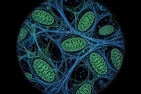
- The team demonstrated it on a human bone cancer cell line, watching mitochondria change shape over time.
- Scientists have developed a new microscopy method that shows the smallest structures inside cells and how their molecular surroundings change, Phys.org reports.
- Researchers from Bielefeld University and the University of Vienna combined two techniques, structured illumination microscopy and fluorescence lifetime imaging, into a method called FLIM-SIM that offers high resolution, fast imaging and a wide field of view.
- The team demonstrated it on a human bone cancer cell line, watching mitochondria change shape over time.
- Scientists have developed a new microscopy method that shows the smallest structures inside cells and how their molecular surroundings change, Phys.org reports.
- Researchers from Bielefeld University and the University of Vienna combined two techniques, structured illumination microscopy and fluorescence lifetime imaging, into a method called FLIM-SIM that offers high resolution, fast imaging and a wide field of view.
- The team demonstrated it on a human bone cancer cell line, watching mitochondria change shape over time.
Scientists have developed a new microscopy method that shows the smallest structures inside cells and how their molecular surroundings change, Phys.org reports. Researchers from Bielefeld University and the University of Vienna combined two techniques, structured illumination microscopy and fluorescence lifetime imaging, into a method called FLIM-SIM that offers high resolution, fast imaging and a wide field of view. The team demonstrated it on a human bone cancer cell line, watching mitochondria change shape over time. The results, part of the European CHEM-SCAN project, were published in the journal Optica.
Scientists have developed a new microscopy method that shows the smallest structures inside cells and how their molecular surroundings change, Phys.org reports. Researchers from Bielefeld University and the University of Vienna combined two techniques, structured illumination microscopy and fluorescence lifetime imaging, into a method called FLIM-SIM that offers high resolution, fast imaging and a wide field of view. The team demonstrated it on a human bone cancer cell line, watching mitochondria change shape over time. The results, part of the European CHEM-SCAN project, were published in the journal Optica.
Scientists have developed a new microscopy method that shows the smallest structures inside cells and how their molecular surroundings change, Phys.org reports. Researchers from Bielefeld University and the University of Vienna combined two techniques, structured illumination microscopy and fluorescence lifetime imaging, into a method called FLIM-SIM that offers high resolution, fast imaging and a wide field of view. The team demonstrated it on a human bone cancer cell line, watching mitochondria change shape over time. The results, part of the European CHEM-SCAN project, were published in the journal Optica.
Most read
سب سے زیادہ پڑھی گئی
सबसे ज़्यादा पढ़ी गई
- Loading...
Get news in your inbox
خبریں اپنے ان باکس میں حاصل کریں
खबरें अपने इनबॉक्स में पाएं
One email when new stories are published. No spam, unsubscribe anytime.
نئی خبریں شائع ہونے پر ایک ای میل۔ کوئی سپیم نہیں، کسی بھی وقت ان سبسکرائب کریں۔
नई खबरें प्रकाशित होने पर एक ईमेल। कोई स्पैम नहीं, कभी भी अनसब्सक्राइब करें।